
A live 500 pound heifer was submitted from a group of feetlot heifers. The animals had been purchased three weeks before at a salebarn.
Picture 1. swollen feet
The problem did not respond to antibiotic treatment, and the lameness progressed to recumbancy. Three other animals at the feedlot were down with a similar history. The toe tips were abraded and oozed foul smelling, black fluid when squeezed.

Picture 2. pedal osteitis
Lateral claws of the hind feet were split which revealed pedal osteitis at the tip of P3 and a cavitated area undermining the sole and hoof wall.

Picture 3. hock joint
A hock joint contained inspissated suppurative exudate.

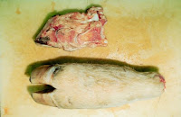
Picture 4. Lungs
Lungs had multiple necrotic lobules, some with large cavitation. Bacterial cultures identified Actinomyces pyogenes and Bacteroides sp. from lung and joint swabs. Pasteurella multocida was also identified from lung and Mycoplasma arginini from joint. This syndrome is known as toe abscess and can occur in cattle handled on rough surfaces. The claw tips are worn until there is white line separation which then allows penetration of dirt and manure into the claw. Bacterial infection with various aerobic and anaerobic microbes results in pedal osteitis, ascending foot infection, and bacteremia. The problem must be identified as soon as possible for treatment to be successful.

Picture 5. interdigital space
The interdigital space is usually not affected, as in footrot. Recommended treatment is trimming the toe to allow drainage (excessive trimming will cause increased lameness) and broad spectrum antibiotic therapy. Some veterinarians will apply a block to the unaffected claw.


No comments:
Post a Comment